

Bradycardia represents a heterogeneous group of sinus node and atrioventricular conduction disorders resulting from impaired impulse formation and/or propagation. Clinical relevance is determined by the underlying substrate, degree of conduction system disease, and associated hemodynamic impact, with advanced bradyarrhythmias contributing to syncope, heart failure syndromes, and malignant bradyasystolic events.

BRADYCARDIA
Bradycardia most commonly reflects sinus node dysfunction or atrioventricular conduction disease, frequently on a substrate of progressive age-related fibrotic degeneration of the conduction system. Risk stratification is driven by conduction disease severity and comorbidity burden, with permanent pacing indicated in clinically relevant cases.
Contemporary device therapy has increasingly incorporated conduction system pacing strategies, including His bundle and left bundle branch area pacing, as physiologic alternatives to conventional right ventricular apical pacing.
Sinus node dysfunction (SND) may be intrinsic or related to drugs, metabolic disorders, hypervagotonia, or carotid sinus hypersensitivity. SND, also known as sick sinus syndrome, encompasses a number of electrocardiographic (ECG) and clinical findings that suggest abnormal impulse generation from the sinoatrial (SA) node.
These ECG findings include: sinus pause, sinus bradycardia (less than 60 bpm), or failure of the SA node to increase its firing rate relative to the physiologic body demand, also known as chronotropic incompetence.
In patients with atrial fibrillation or other tachyarrhythmias, sinus node disease manifests with the so-called “tachy-brady syndrome”.
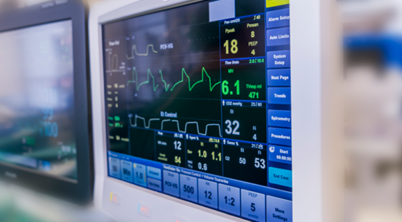
While SA node disease is a problem of impulse generation, atrioventricular (AV) conduction disease is one of impulse propagation. This manifests electrocardiographically with conduction delay or conduction block and results in bradycardia and pauses.
TYPES OF ATRIOVENTICULAR BLOCKS
FIRST-DEGREE ATRIOVENTRICULAR BLOCK
This is defined as a lag between the time that the SA node fires and actual depolarization of the atria. This rhythm is not recognizable on an ECG strip because a strip does not denote when the SA node fires.
SECOND-DEGREE ATRIOVENTRICULAR BLOCK
Second degree SA blocks are broken down into two subcategories just like AV blocks are:
- Second degree type I (Wenckebach block): an irregular rhythm, and an R-R interval that gets progressively smaller, while the P-R interval remains constant, until a QRS segment is dropped
- Second degree type II (sinus exit block): A regular rhythm that may be normal or slow. It is followed by a pause that is a multiple of the P-P interval usually2-4. Conduction across the SA node is normal until the time of the pause when it is blocked.
THIRD-DEGREE ATRIOVENTRICULAR BLOCK
Third degree block is caused by failure to conduct them. The rhythm is irregular and either normal or slow. It is followed by a long pause that is not a multiple of the P-R interval. The pause ends with a P wave, instead of a junctional escape beat the way a sinus arrest would.
TV
TV

Understanding AVEIR VR Retrieval

Why Leadless Pacing Summit 2026 Matters

AVEIR Atrial LP AAI Implant Training Supplement
AVEIR DR Virtual Implant Training Supplement
Leadless in Congenital Patients
When Patients Evolve: Upgradability in Leadless

Optimizing Tricuspid Valve Crossing: Case example

Optimizing Tricuspid Valve Crossing: Case example

Health Economics of Leadless Therapy
No Wait MRI, an Assurity MRI™ patient story
- NHS. Heart rhythm problems (arrhythmia). Available at: www.nhs.uk/conditions/arrhythmia/. Accessed June 11, 2026.
- American Heart Association. Tachycardia: Fast heart rate. Available at: www.heart.org/en/health-topics/arrhythmia/about-arrhythmia/tachycardia--fast-heart-rate. Accessed June 11, 2026.
- Mayo Clinic. Supraventricular tachycardia – Symptoms and causes. Available at: www.mayoclinic.org/diseases-conditions/supraventricular-tachycardia/symptomscauses/ syc-20355243. Accessed June 11, 2026.
- Mayo Clinic. Ventricular tachycardia – Symptoms and causes. Available at: www.mayoclinic.org/diseases-conditions/ventricular-tachycardia/symptoms-causes/syc- 20355138. Accessed June 11, 2026.
- Cleveland Clinic. Sinus tachycardia: Causes, symptoms & treatment. Available at: my.clevelandclinic.org/health/diseases/23210-sinus-tachycardia. Accessed June 11, 2026.
- Issa Z, Miller J, Zipes D. (2012). Clinical Arrhythmology and Electrophysiology, 212–238. Philadelphia, PA: Saunders Elsevier.