

Bradycardia represents a heterogeneous group of sinus node and atrioventricular conduction disorders resulting from impaired impulse formation and/or propagation. Clinical relevance is determined by the underlying substrate, degree of conduction system disease, and associated hemodynamic impact, with advanced bradyarrhythmias contributing to syncope, heart failure syndromes, and malignant bradyasystolic events.
DIAGNOSING BRADYCARDIA
A careful history and physical examination are essential for the evaluation of patients with suspected or documented bradycardia.
Current guidelines emphasize the importance of the history and physical examination in the initial evaluation, particularly for identifying patients with structural heart disease.1 Together with the history and physical examination, the resting electrocardiogram (ECG) is an essential component of the initial evaluation of patients with documented or suspected bradycardia.1
LEARN MORE:

SYMPTOMS MAY INCLUDE:2

Chest pain

Dizziness or lightheadedness

Fainting or near-fainting (syncope)

Confusion or memory problems

Feeling very tired, especially during physical activity

Shortness of breath
When patients present with symptoms of bradycardia or pauses such as syncope, lightheadedness, fatigue, loss of energy or stamina, and the 12-lead ECG is not diagnostic, ambulatory monitoring is often considered. Types of monitoring include:

Holter monitors
Automatic trigger or patient-activated monitors

Insertable cardiac monitors
Holter monitors are used with daily symptoms. Automatic trigger or patient-activated monitors can be used for weeks at a time when symptoms are less frequent. For even more rare symptoms, especially with syncope, implanted loop recorders are sometimes used; these can extend the monitoring period for up to 6 years.
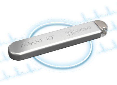
The Assert-IQ™ insertable cardiac monitor (ICM) is indicated for the monitoring and diagnostic evaluation of patients who experience unexplained symptoms that may be cardiac-related such as: dizziness, palpitations, chest pain, syncope, and shortness of breath, as well as patients who are at risk for cardiac arrhythmias such as bradycardia, tachycardia, and sinus pauses.
In the case of presyncope or syncope, where there is no documented bradycardia or heart block on ambulatory monitoring, yet suspicion for sinus node dysfunction (SND) or atrioventricular (AV) conduction disease remains high, invasive electrophysiologic testing is performed.
SA node function is studied by first pacing the atrium at a specified rate. Pacing is then terminated, and the time from the last pacing impulse to the first sinus beat is measured, which is called the sinus node recovery time (SNRT). The corrected SNRT is the difference of the SNRT and the pacing cycle length.
A value greater than 525 milliseconds is considered abnormal.

Certain genetic mutations have been linked to sinus node and conduction system disease. SCN5A, HCN4, RYR2, CASQ2, and ankyrin-B (ANKB) mutations are associated with SND, whereas mutations of SN5A, SCN1B, KCNJ2, TBX5, and NKX2-5 are associated with conduction system disease.
Neuromuscular genetic disorders including emerin (EMD), lamin A/C (LMNA), and myotonic dystrophy type 1 (DM1) are also associated with AV conduction disease.
Hub
TV

Understanding AVEIR VR Retrieval

Why Leadless Pacing Summit 2026 Matters
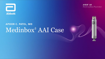
AVEIR Atrial LP AAI Implant Training Supplement
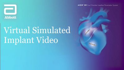
AVEIR DR Virtual Implant Training Supplement
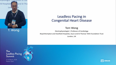
Leadless in Congenital Patients
When Patients Evolve: Upgradability in Leadless

Optimizing Tricuspid Valve Crossing: Case example

Optimizing Tricuspid Valve Crossing: Case example

Health Economics of Leadless Therapy
No Wait MRI, an Assurity MRI™ patient story
- Glikson M, Nielsen JC, Kronborg MB, et al. 2021 ESC Guidelines on cardiac pacing and cardiac resynchronization therapy. Eur Heart J. 2021;42(35):3427-3520. doi: 10.1093/eurheartj/ehab364. Erratum in: Eur Heart J. 2022 May 1;43(17):1651. doi: 10.1093/eurheartj/ehac075.
- Mayo Clinic. Bradycardia Overview. Available at: www.mayoclinic.org/diseases-conditions/bradycardia/symptoms-causes/syc-20355474. Accessed June 23, 2026.